In this issue:
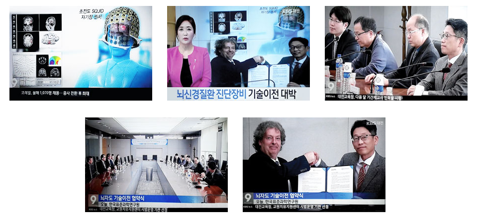
South Korean news channel KBS reported Compumedics’ and South Korea’s KRISS (Korea Research Institute of Standards and Science) MOU deal covering immediate arrangements for the MEG superconducting quantum interference device and clean room fabrication. This enables immediate orders, sales and manufacturing arrangements for Compumedics’ new MEG (MagnetoEncephaloGraphy) systems. The MEG systems are expected to be priced from $4m to $6m per system.

The CURRY NeuroImaging platform and MEG have a history stretching back over 25 years. CURRY was first conceived as a product in the early 1990’s when Philips Electronics investigated the feasibility of developing it’s own MEG hardware platform. Ultimately, the hardware platform did not survive, but the software, along with its core engineering architects, Dr. Manfred Fuchs & Dr. Michael Wagner, continued on. When Philips exited the MEG business, CURRY and the development team were purchased by Neuroscan. At this time, the UNIX-based CURRY platform appealed more to the research community than to the clinical market. By 1999, publications were emerging describing the application of CURRY for cortical localization of EEG and MEG activity for tactile and auditory sensory input. However, “novel developments” and “new approaches to detailed localization of specific epileptic discharges” as well as identification of functionally critical areas of the brain controlling language and memory using CURRY, were also being reported in the clinical literature. Migration of CURRY from the UNIX to Windows platform in 2003 resulted in a rapid expansion of the use of CURRY in both the research and clinical worlds.
The benefits associated with CURRY’s ability to integrate MEG with EEG and co-register both kinds of high temporal resolution functional imaging data with the structural neuroimaging data including MRI, CT, DTI, as well as PET, SPECT and fMRI accelerated the the adoption of the software by both the research and clinical communities. Early clinical adopters, such as Dr. John Ebersole, supported and championed the benefits of source localization tools such as CURRY, contributing to the development of specific source analysis billing codes for EEG and MEG. For a long time, CURRY has been the de-facto software platform for clinical MEG community, particularly for those assessing epilepsy. This has culminated in the adoption of CURRY as the standard analysis platform by the European Epilepsy Consortium. For the CURRY team, integrating CURRY with the KRISS MEG hardware represents a full circle of development. With long-term future development plans for both hardware and software, CURRY MEG will offer an expanding list of benefits based on the first fully integrated platform combining EEG, MEG, multi-modal neuroimage co-registration and source reconstruction from a single provider – Compumedics Neuroscan.
To facilitate the commercial success of the CURRY MEG system, Compumedics Neuroscan is pleased to welcome Mr. Stephen Otto, former Chairman & Chief Operating Officer of Elekta Neuromag, who will provide Sales and Marketing support as we move forward into this new market.

We’ve signed a major exclusive deal with Korea’s equivalent of Australia’s CSIRO, which has the potential to advance the global neuro-function imaging market and transform Compumedics/Neuroscan (CMP/NSC) business.
The licence and technology transfer agreement with the KRISS team was signed and announced to the ASX last month, providing access to state-of-the-art brain imaging technology called magneto-encephalography or MEG for short.
The deal brings together the Korean Institute of Standards and Science (KRISS) new generation radial gradiometer (versus earlier generation and less sensitive planar magnetometer) neuroimaging system with CMP/NSC market-leading brain analysis software.
The significant imaging market opportunities this will open have the potential to generate more than US$20 million in incremental annual CMP/NSC revenues in about two years.
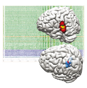
MEG enables a superior neuro-functional view of the brain by way of identifying and tracking sensitive magnetic field activity, which has greater sensitivity and gives faster measurement than other imaging technologies. This will help in the early diagnosis and management of some of the most prevalent and debilitating neurological disorders including Alzheimer’s, Epilepsy, Parkinson’s and Autism, as well as traumatic brain injury. When recorded and analysed with Compumedics CURRY Neuroimaging Suite, EEG may be acquired simultaneously, providing additional inf ormation about the origins of disrupted brain activity. When recorded in isolation, MEG has the immense advantage of not requiring any special preparation; the patient’s head is simply placed inside the helmet containing the MEG electronics, and the magnetic fields are obtained without any contact. Therefore, as well as adults it is also ideally suited for pediatric neurology, which is the most critical expanding patient-population for surgical remediation of epilepsy.
The deal is a key milestone in our strategy to transition from being a technology leader in the US$50 million specialist brain research market to the much larger US$4 billion multi-modal brain imaging market.
We will change from selling US$30,000 MEG software packages to selling best of class fully integrated US$5.5 million MEG systems.
The technology we’ve acquired is complementary to MRI (magnetic resonance imaging) or PET (positron emission tomography) scan but provides 50 per cent greater spatial resolution than the major MEG competitor currently on the market. That is how it gives a better view of the brain and its activity. This technology expands our capability in multi-modal brain analysis, where the clinician wants to combine two or more imaging modes – such as electroencephalography (EEG), MRI and MEG – to locate and assess brain electrical activity non-invasively with unprecedented accuracy.
> The agreement with KRISS is important because its technology will go to the market only with our software as well as our EEG hardware for the dual imaging systems.
> By financial year 2018-19, we expect to be selling US$20 million a year worth of complete new-generation MEG brain imaging systems.
> The early detection of neurological disorders is a huge advantage because many are treatable if discovered early.
> Not only can this save lives, it can also save government dollars. Given the ageing populations in much of the developed world – and also in China and India – health spending is mushrooming, which means governments have to choose the most effective diagnostic methods and treatments.
> In particular, China and India are starting to spend heavily on their healthcare infrastructure and they don’t want to go through the intermediate brain-imaging technologies that the developed world went through. They want to go straight to state-of-the-art systems.
> Demand for brain imaging services is growing much faster than health spending in general.
In this fast-growing market, we’ve hit the sweet spot at the right time. For us, brain imaging is now our third globally significant technology platforms behind premium sleep diagnosis systems and neurology/blood flow diagnosis systems. In time we see it generating most of our new sales.
Invented by David Cohen at Massachusetts Institute of Technology in the 1970s, MEG technology can record the magnetic fields produced by electric currents generated by neurons in the brain.
It does this using a highly sensitive detector called a SQUID, short for super-conducting quantum interference device.
By maintaining superconducting magnetic detectors at super-cooled temperatures, the tiny but very fast (too fast for traditional MRI) brain characteristics associated with widespread neurological disorders such as Dementia, Autism, Epilepsy
can be detected.


