CURRY®
Signal Processing and Source Localization Multi-Modal Neuroimaging Suite
The history of the CURRY® software is well known. Throughout the world, CURRY® has the well deserved reputation as the most advanced and comprehensive tool for Multimodal Neuroimaging. CURRY’s strength has historically been combining functional data such as EEG and MEG with structural data from MRI and CT to optimize source reconstruction.
Compumedics Neuroscan’s CURRY® software is FDA-cleared and CE-marked.
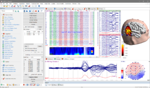
It features a complete set of tools for efficient EEG/MEG/ERP recording, review, and multi-modal integration – presented in a modern and clean user interface.
Over three decades of research and collaboration with leading brain research laboratories and Epilepsy units around the
world have resulted in proven features and functionality that continue to advance the field.
For further information on this product, please visit www.compumedicsneuroscan.com
Explore some of the product highlights below.
Compumedics Neuroscan CURRY® One software, modular architecture
- Record EEG, ERP, MEG, fMRI-EEG from Compumedics Neuroscan USB, network, and wireless amplifiers
- Review and process data recorded on Compumedicsor third-party systems in time, space, and spectral domain
- Integrate with MRI, CT, PET, SPECT, fMRI, DTI
- Localize brain activity in individual head and brain anatomy
- Visualize results and integrate modalities in an interactive 3D environment
- Available configurations range from from small lab edition (for up to 48 EEG channels and 4 kHz sampling rate) to fully featured clinical epilepsy analysis (up to 512 EEG channels and 20 kHz sampling rate).
Data Acquisition with Online Processing
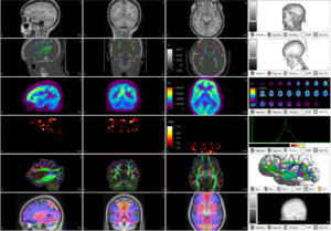
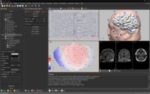
Multiple Co-registered Image Data Sets
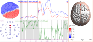
Statistical Analysis
CURRY® for Epilepsy Applications
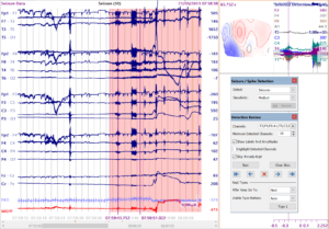
- Combined review of waveform data and scalp topographies
- Automatic spike and seizure detection based on machine learning
- Accurate manual marking of spikes and interactive spike type editor
- Search for further occurences of manually marked spikes
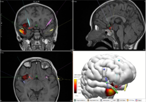
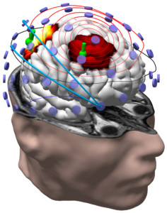
- Source localization of spikes and seizure onsets
- Dipole clustering shows localization variability of spikes per type
- Cortical localization of spike onset using extended patches of inward-flowing currents shows candidate generating gray matter
- Automatic co-registration and overlay with MRI, CT, PET, SPECT, DTI
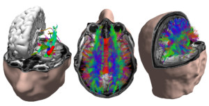
- DTI fiber tracking for exploring connectivity between foci
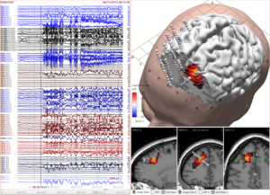
- Intracranial stereo-EEG, grid and strip electrode placement planning and export to surgical navigation systems and robots
- Intracranial electrode contact localization from post-implant CT or MRI
- Review of intracranial data in anatomical context
- HFO analysis helps identifying contacts-of-interest
- Source localization of intracranial data for foci not covered by contacts
- FDA-cleared and CE-marked.
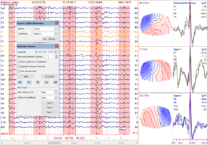
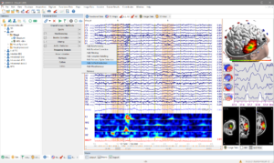
Spike Detection and Review
Seizure Detection and Review
Stereo-EEG Planning
DTI Fiber Tracking
Stereo-EEG Review and Source Localization
Optimized for Efficient Workflow
- Adaptive user interface helps focus on the tasks at hand by hiding currently unused options
- Scopes manage application area-dependent presets
- Flexible data display shows single or combined aspects such as waveform views, spectral and time-frequency displays, topography maps, image data cross-sections, and 3D renderings
- Workflow manager shows open tasks, presents user
interfaces and data displays to get things done quickly - Reporting tool collects images and text from ongoing analysis
- 64-bit software architecture with multi-core optimization and unlimited memory access for highest performance standards.
Signal Processing and Source Localization
Dark Mode
Automation
- Macro Recorder captures and replays simple or complex processing sequences, no programming skills required
- Batch processing applies recorded macros to multiple studies.
Sensor Coherence

